See This Report on Babyecho
See This Report on Babyecho
Blog Article
The Greatest Guide To Babyecho
Table of ContentsBabyecho Can Be Fun For EveryoneThe Definitive Guide to BabyechoBabyecho Can Be Fun For EveryoneThe Buzz on BabyechoThe Only Guide to BabyechoThe Ultimate Guide To BabyechoThe smart Trick of Babyecho That Nobody is DiscussingThe Buzz on BabyechoBabyecho - Truths
You may not listen to the baby's heart beat at the very first trimester ultrasound consultation, which's fine. It might be since you're not much enough along in the maternity or the child's position. Often the infant's heart beat can be detected as early as 5 to 6 weeks after perception. You can typically listen to the heart beat much better more detailed to 10 weeks after gestation.:max_bytes(150000):strip_icc()/191127-ultrasound-trimester-pink-2000-fd089add04f8444e9d7a403933d1994f.jpg)
The Best Guide To Babyecho
An ultrasound additionally called a sonogram assists your medical professional figure out whether the fetus is creating normally. Your doctor may advise that you have 1 or even more ultrasounds at different points in your pregnancy. It can be made use of to check the composition of the fetus for flaws or troubles. Depending upon just how far along your maternity is, ultrasound photos help your medical professional: estimate your due date check things like the size and placement of the unborn child to ensure everything is normal see the position of the placenta see the amount of amniotic liquid in your womb locate numerous pregnancies (twins, triplets, etc) Ultrasounds can additionally be used to screen for sure abnormality, like Down syndrome.
Medical professionals, midwives, or educated ultrasound professionals will do your ultrasound and review the outcomes. The price of an ultrasound depends on the type of ultrasound you obtain and where you get it.
Babyecho for Beginners
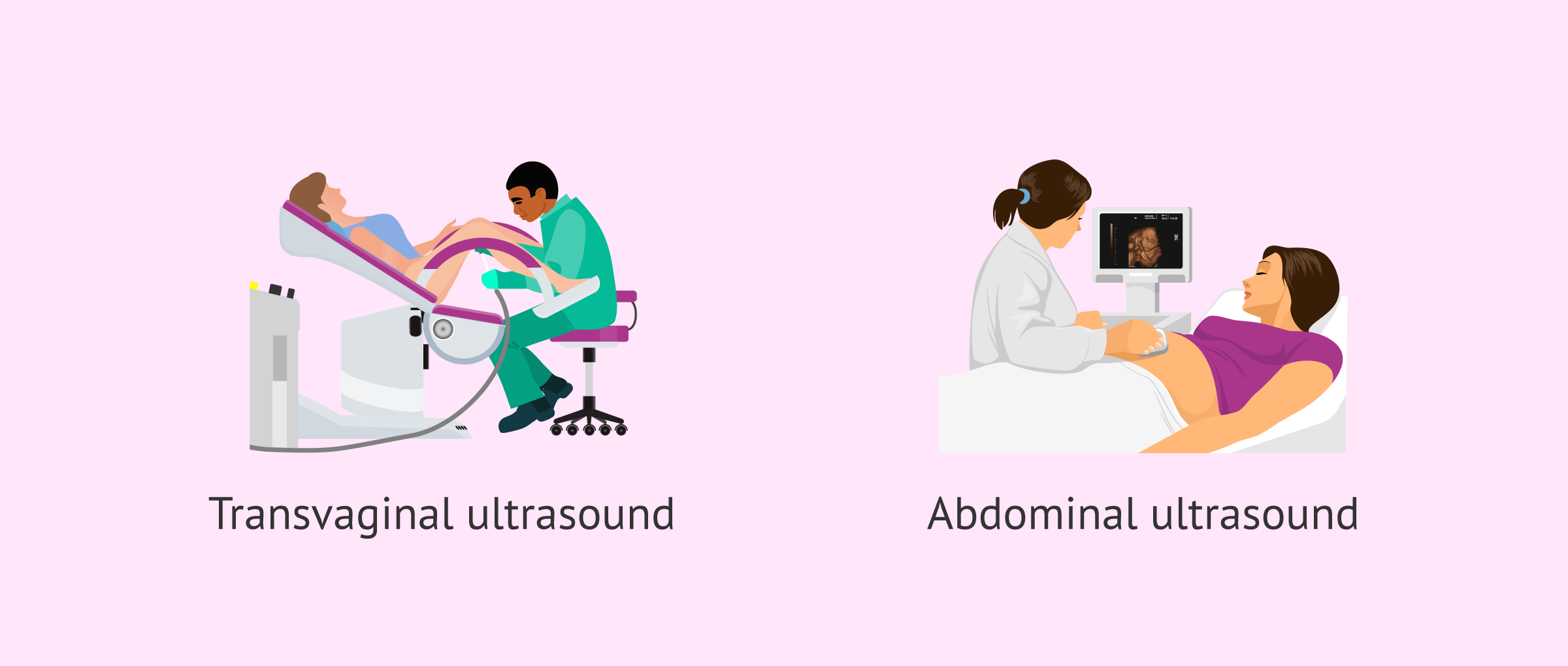
Ultrasound scans make use of sound waves to develop an image of your baby on a TV display. Most ultrasounds are 'transabdominal ultrasounds'.
You will be brought into a darkened room. The darkness makes it less complicated for the individual doing the check to plainly see the picture on the display. You will certainly be asked to exist on a couch and raise your top to reveal your stomach. You might require to roll down the waistband of your trousers.
3 Easy Facts About Babyecho Described
This is to shield your clothing from the ultrasound gel. Next, they will certainly put the ultrasound gel onto your tummy. The gel aids the probe to move. As the probe conforms your stomach, an image will appear on the screen. The pictures you see remain in black and white.
You might not be able to tell where your child remains in the picture. It is regular for the images to look a little gloomy or blurry in the beginning. The individual doing the check will often aim things out to you like the baby's heart beat and head. The check does not harmed.
Everything about Babyecho
It can be upsetting when your check suggests that there is an issue with your pregnancy or your baby. Your midwife, obstetrician and General practitioner are there to sustain you.
They can be costly and you will certainly require to reserve the scan on your own. Your GP, midwife or obstetrician will certainly tell you where you can get exclusive pregnancy scans click site in your location. If a concern is found on a personal check, follow-on solutions might not be readily available there. You will certainly require to contact your maternity health center.
Fetal ultrasound is a test done throughout pregnancy that uses shown sound waves. The image is shown on a Television screen. It might be in black and white or in colour.
See This Report about Babyecho
It can be done as early as the Fifth week of pregnancy. Sometimes the sex of your unborn child can be seen by regarding the 18th week of pregnancy.
Estimate the age of the unborn child (gestational age). Estimate the danger of a chromosome flaw, such as Down disorder. Look for birth flaws that impact the brain or spinal cord. This examination is done to: Estimate the age of the fetus. Check out the size and placement of the unborn child, placenta, and amniotic liquid.
The 6-Minute Rule for Babyecho
It can be distressing when your check suggests that there is a trouble with your maternity or your infant. Your midwife, obstetrician and general practitioner exist to sustain you. Ask to discuss everything to you thoroughly. You might need extra scans if: you have bleedingyou have concerns regarding your baby's movementsyour child's development requires to be monitoredExtra scans might also be needed to inspect: the placement your baby is existing inthe position of the placenta (afterbirth)the quantity of amniotic fluid around your babyMost pregnancy hospitals do not routinely do scans to determine the sex of your child.

About Babyecho
Fetal ultrasound is an examination done during pregnancy that uses reflected audio waves. The picture is displayed on a TV display. It may be in black and white or in colour.
It can be done as early as the 5th week of pregnancy. Often the sex of your unborn child can be seen by about the 18th week of pregnancy.
Babyecho Fundamentals Explained
Inspect for birth problems that influence the mind or back cable. This test is done to: Estimate the age of the unborn child. Look at the size and position of the fetus, placenta, and amniotic fluid - doppler ultrasound.
Report this page